Phacoemulsification
This is the latest technique of cataract surgery. Here the cataract is operated through a 3.2 - 5 mm wound, which doesn't require any suture application. After emulsifying the cataractous lens by phacoemulsification probe based on ultrasonic vibration, artificial intraocular lens is implanted. The intraocular lens used can be foldable or non-foldable type. The foldable lens implantation requires a smaller incision as compared to non-foldable one. Intraocular Lens Implantation - Various IOLs implanted are Non-foldable, Foldable, Blue blocking, Voilet shield, Multifocal, Toric IOLs ete.
Glaucoma Clinic
- Tonometry
- Automated Perimetry
- HRT-II
- Gonioscopy
- Glaucoma Surgery
At our centre we have the facilities of diagnosing and treating the patients of glaucoma.  Glaucoma is a disease in which the pressure inside the eye rises. This raised pressure tends to damage the nerve of the eye (optic nerve). We have the facilities of Intraocular pressure recording by computerized and non-computerized methods both.
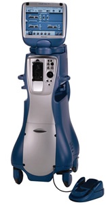
It is a newer diagnostic modality in Glaucoma. By this computerized equipment, optic nerve structure is analyzed so as to know the glaucomatous changes at an earlier stage. The field charting of glaucoma patients is done by automated perimeter (Humphry). The angle of anterior chamber of eye is assessed by Gonioscopy. The contrast sensitivity is assessed by Cambridge contrast sensitivity chart. Those patients of glaucoma who are not controlled by medicines are taken up for glaucoma surgery. Those patients whose examination shows their proneness for attacks of glaucoma are subjected to YAG laser iridotomy.
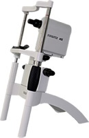
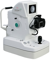
The patients having retinal diseases are subjected to fundus photography so that the retinal lesion is under observation. The photographs are taken by fundus camera. Fundus Fluorescein Angiography The patients of diabetic retinopathy, age related macular degeneration, maculare oedema and certain other conditions where the leakage through the vessels is doubted, are subjected to fluorescein angiography. In this procedure, we inject the fluorescein dye through the blood vessel of arm and take the fundus photographs. This procedure tells us about the possible leaking vessels and accordingly the patient is taken up for laser photocoagulation or intravitreal injections of drugs.
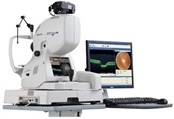
Spectral Domain OCT This is ultramodern computerized equipment which is used to scan retina & optic nerve. In this technique retinal layers are analyzed to diagnose a disease by non-touch technique and without using any injection. By OCT, macular ocdema (swelling in center of retina) is measured and then compared after treating the patient.
The patients having retinal diseases are subjected to fundus photography so that the retinal lesion is under observation. The photographs are taken by fundus camera. > Retinal Detachment > Posterior Vitrectomy > Pneumoretinopexy > Retinal Detachment + Vitrectomy + > Silicone Oil Injection > Macular hole surgery
PASCAL Laser
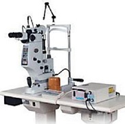
Pattern scan laser photocoagulation is a fully integrated system designed to treat retinal diseases using a single spot or a predetermind pattern array of upto 56 spots. PASCAL represents a significant leap forward from single spot green laser photocoagulation. In conventional laser, one foot pedal depression leads to one shot to the retina while in PASCAL laser with a single depression of foot switch multiple spots can be applied almost simultaneously. What are the benefits of PASCAL? > The speed > Improved comfort > Reduce the number of the laser sessions > Advanced Precision > Lesser Adverse Effects
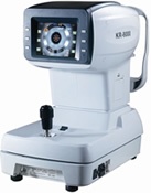
This facilitates calculation of power of lens required to correct the refractive error.
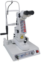
After Cataract surgery, many a time patients tend to have thickening of posterior capsule (Posterior capsule is the membrane left behind during cataract surgery for the support of intra-ocular lens). Because of the opacified posterior capsule, the patient's vision deteriorates. By YAG laser, an opening is made in the opacified posterior capsule and thus the visual acuity of patient is restored. YAG laser is also used to make an opening in the iris so that the possibility of acute congestive glaucoma is lessened.
We also offer the facility of surgical intervention in cases of squint.
We have also the facility of operating the patients of entropion, ectropion, ptosis etc.
A Scan: By this the axial length of eyeball is measured and the interaocular lens power is calculated. This facilitates calculation of power of lens required to correct the refractive error. B Scan: This equipment is used to assess the vitreous (Gel like structure in front of retina) and retinal when these two are not visible clinically because of dence calaract or any other reason for hazy media.
Here, we are prescribing contact lenses and the lenses available with use are soft, toric, therapentic C.L. and cosmetic C.L.


